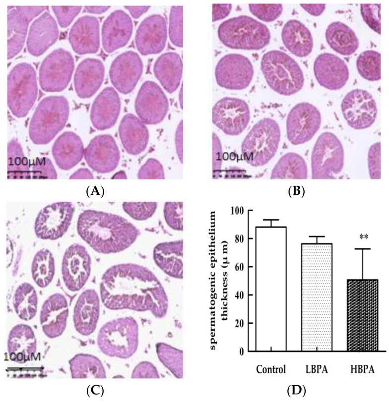
Figure 5.
Effects of perinatal BPA exposure on pathological damage in the testis tissue of male offspring. Histology of control mice (A), 0.2 μg/mL BPA (B), and 2 μg/mL BPA (C). Semi-quantitative analysis of testicular spermatogenic epithelium thickness by hematoxylin and eosin (H&E) staining (D). ** p < 0.01 compared with controls. Magnification: A–C, 200×.

