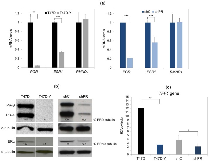
Figure 1.
Loss of PR reduces the ESR1 expression in hormone-deprived T47D breast cancer cells. (a) Gene-specific mRNA expression measured by quantitative RT-PCR in T47D or PR-deficient cells (T47D-Y) (left panel) and T47D cells transduced with shRNA against PR (shPR, clone trcn0000010776) or scramble shRNA (shC) (right panel). The gene-specific expression levels were normalized to GAPDH expression and are represented as relative values in the T47D cells. RMND1 was used as a PR-independent control. PGR, PR-encoding gene; ESR1, ER-encoding gene. Error bars represent the SD of three independent experiments. ** p less than or equal to 0.01, *** p less than or equal to 0.005, unpaired two-tailed Student’s t-test. (b) PR and ERα protein levels measured by Western blot in T47D and T47D-Y cells (left panel) and in T47D transduced with shRNA against PR (shPR; clone trcn0000010776) or scramble shRNA (shC) (right panel). α-tubulin protein was used as the loading control. The intensities of the PR and ER bands were normalized to α-tubulin and represented as the relative value in the control cells. The vertical white line depicts a removed lane between the two samples. Blots are representative of three independent experiments. (c) PR depletion impairs TFF1 estrogen-mediated gene transcription. T47D cells, PR-deficient (T47D-Y) cells, short hairpin control (shC) and PR-depleted cells (shPR, clone trcn0000010776) were treated with estradiol (E2, 10 nM) or ethanol (vehicle) for 6 h, at which point TFF1 mRNA expression was measured by quantitative RT-PCR. The TFF1 gene expression was normalized to GAPDH expression and is represented as fold change relative to the vehicle (E2/vehicle). Error bars represent the standard deviation (SD) of three independent experiments. * p less than or equal to 0.05, ** p less than or equal to 0.01, unpaired two-tailed Student’s t-test.

