Figure 3.
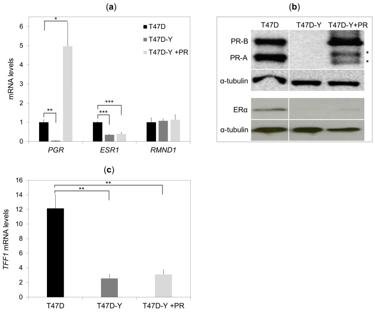
PR rescue of PR-deficient cells does not restore ESR1 gene expression. (a) Gene-specific mRNA expression measured by quantitative RT-PCR in T47D control cells, PR-deficient cells (T47D-Y) and PR-rescue cells (T47D-Y + PR). The mRNA expression levels were normalized to GAPDH expression and are represented as values relative to the T47D cells. PGR, PR-encoding gene; ESR1, ER-encoding gene. Error bars represent the SD of three independent experiments. * p less than or equal to 0.05, ** p less than or equal to 0.01, *** p less than or equal to 0.005, unpaired two-tailed Student’s t-test. (b) Gene-specific protein levels measured by Western blotting in T47D control cells, T47D-Y and T47D-Y + PR cells. The lanes T47D and T47D-Y of this image are the same as in Figure 1b and are shown here for comparison with T47DY +PR. Blots are representative of three independent experiments. * indicates the degradation products of PR-B isoform. (c) PR rescue of PR-depleted cells does not restore the estrogen-mediated gene expression. T47D, PR-deficient cells (T47D-Y) and PR-rescue (T47D-Y + PR) cells were treated with estradiol (E2, 10 nM) or ethanol (vehicle) for 6 h, at which point, TFF1 mRNA expression levels were measured by quantitative RT-PCR. Gene-specific expression levels were normalized to GAPDH expression and are represented as values relative to the vehicle (E2/vehicle). Error bars represent the SD of three independent experiments. ** p less than or equal to 0.01, unpaired two-tailed Student’s t-test.

