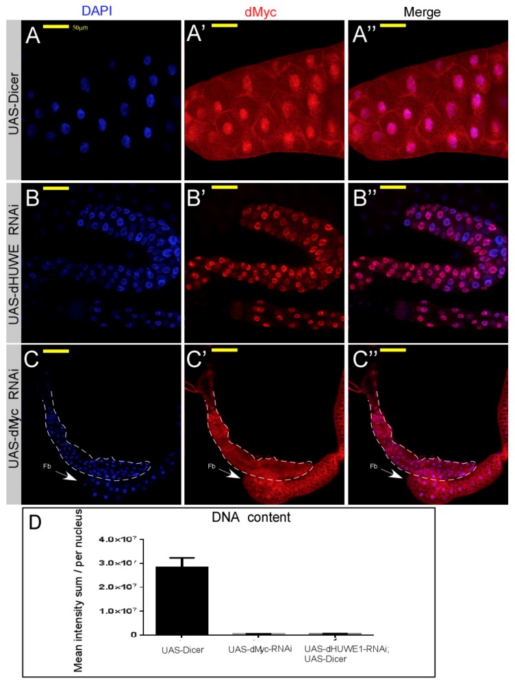
Figure 2.
Loss of dHUWE1 results in a small salivary gland that expresses dMyc protein. (A–C”) Indirect immunofluorescence confocal microscopy images of wild-type (A–A”) and the indicated UAS-RNAi transgenic lines (dHUWE1, B–B”; dMyc, C–C”) immuno-stained with anti-dMyc antibody (red). DAPI (blue) marks DNA, and the scale bar is 50 μm. The dotted line in C–C” indicates the edges of the small salivary gland. Fb; fat body tissue. (D) Indirect quantification of the DNA content of salivary gland cells derived from the indicated transgenic lines using Imaris technology® (n = 10 p < 0.001).

