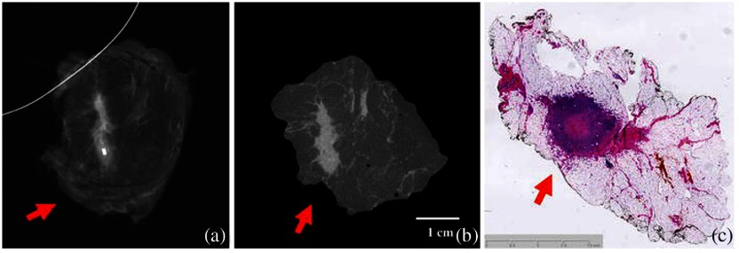
Fig. 2.
(a) Tumor mass appears far from the margin in 2-D radiography. (b) Micro-CT cross section indicates tumor closer to the edge of the specimen. (c) Histopathology slide shows tumor mass closer to edge, more similar to the micro-CT slice than the 2-D radiography image.48 The mammography images are limited to one or two views, while the micro-CT is full volumetric. The whole mount histology is useful but not routinely done for any lumpectomy specimens, so the value of a micro-CT is to visualize the tumor extent in all 3-D. Reprinted by permission from Ref. 48, Springer Nature.

