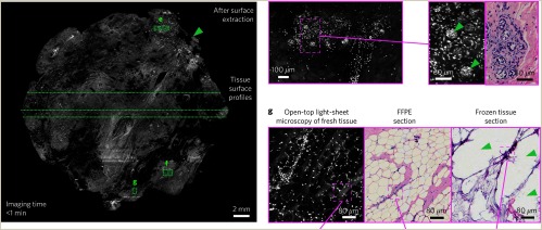
Fig. 8.
Images comparing the performance of light-sheet microscopy with histology. Images of different regions with different magnifications and their corresponding histology images are shown. Results from frozen-sectioning imaging are also included and suggest improved performance with light-sheet microscopy.103 The value of this type of system is the extremely high spatial resolution and the ability to augment pathology imaging, whereas the limitations here are the fact that the scan times could be lengthy and the information could be too dense for surgical use. Reprinted by permission from Ref. 103, Springer Nature.

