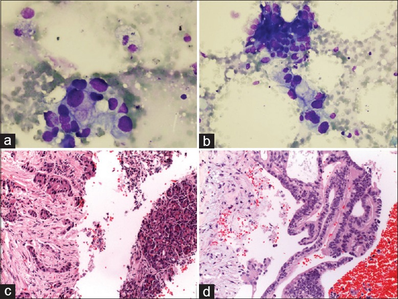
Figure 1.

(a and b) Cytologic feature of PDAC: loosely arranged epithelial cells showing marked nuclear enlargement, nuclear membrane irregularity, anisonucleosis (MGG ×400). (c and d) Cell block sections of a PDAC. (c)Irregular, angulated small tubules are embedded in a sclerotic stroma; in the right part, there is a nontumoral pancreatic tissue. (d) Pseudostratified tumor cells that form fused glands and cribriform structure (H&E ×400)
