
Figure 1.
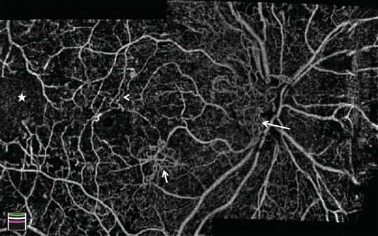
Montage optical coherence tomography angiography image of the optic disc and macula in a patient with diabetic retinopathy. Vascular changes are shown, including enlarged foveal avascular zone, optic disc neovascularization (long arrow), macular neovascularization (short arrow), microvascular tortuosity (arrowhead), and extensive capillary nonperfusion (star).