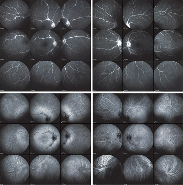
Figure 4.
Fluorescein and indocyanine green angiographic images of a 43-year-old woman with ocular sarcoidosis showing predominant retinal involvement. Fluorescein angiography (intermediate angiographic phase) showing optic disc hyperfluorescence and retinal vascular staining and leakage in both eyes (top right and left). The fluorescein angiographic score was 7 in the right eye and 6.5 in the left eye. Indocyanine green angiography (late phase) showing both eyes (bottom right and left) had small hypofluorescent dark areas along the retinal vessel producing a mask effect. The indocyanine green angiographic score was 2 in both eyes.

