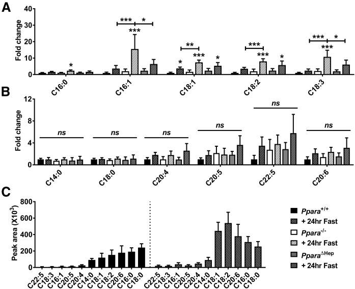
Fig. 5.
Extrahepatic PPARA normalizes serum FA response in fasted PparaΔHep mice. Mice were fed ad libitum or fasted for 24 h, and then serum was collected for analysis. Ppara-dependent (A) and -independent (B) changes in serum FAs in fasted Ppara+/+, full-body Ppara-null (Ppara−/−), and hepatocyte-specific Ppara-null (PparaΔHep) mice. C: Mean MS peak area data indicating relative abundance of individual FAs in fed and fasted WT (Ppara+/+) mouse serum. Fold change data are shown as mean fold change ± SD (n = 5) versus fed WT groups. Asterisks above bars indicate significance versus fed group of the same genotype. * P < 0.05; ** P < 0.01; *** P < 0.001; ns, not significant.

