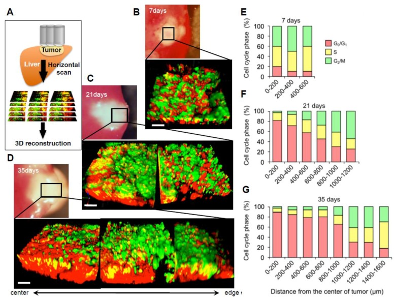
Figure 3.
Three-dimensional image of FUCCI-expressing tumor reveals a vast majority of quiescent cancer cells. (A) Schematic diagram of in vivo CLSM imaging of different-sized tumors. Tumors were scanned from the center to the edge. 800 × 800 pixels and 1.0 μm z steps were scanned, which took 1–2 s per section, with 6–8 min per full 3D scan. The tracing data were imported to Velocity 6.0 version (Perkin Elmer), where all further analyses were performed, and then the scanned images were three-dimensionally reconstructed. (B–D) Representative 3D reconstruction images of a nascent tumor at 7 days after cancer-cell implantation (B), 21 days (C), and 35 days (D) after implantation. (E–G) Histograms show the distribution of FUCCI-expressing cells at different distances from the center. The number of cells in each cell-cycle phase was assessed by counting the number of cells of each color at the indicated time points. The percentage of cells in the G2/M, S, and G0/G1 phases of the cell cycle is shown. Scale bars represent 100 μm. (Reproduced from [46] with the permission of Taylor and Francis).

