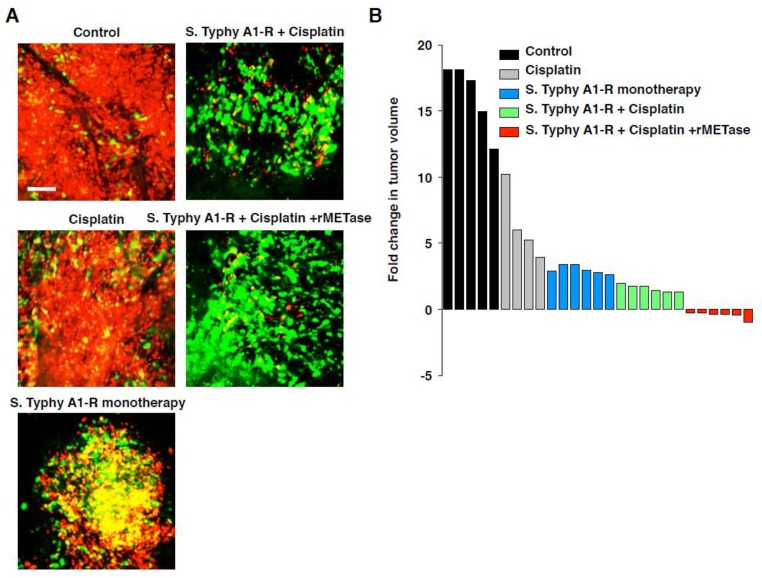
Figure 7.
Decoy, trap, and shoot chemotherapy guided by FUCCI imaging. FUCCI-expressing MKN45 cells (5 × 10⁶ cells/mouse) were injected subcutaneously into the left flank of nude mice. When the tumors reached approximately 8 mm in diameter (tumor volume, 300 mm3), mice were administered S. typhimurium A1-R i.v. alone; or with CDDP (5 mg/kg i.p.) alone for 5 cycles every 7 days, or with a combination of S. typhimurium A1-R and CDDP; or with a combination of S. typhimurium A1-R, rMETase (200 U/day, for 3 days/cycle, 5 cycle), and CDDP (5 mg/kg, i.p.). (A) Representative images of cross-sections of FUCCI-expressing MKN45 tumors; untreated control; S. typhimurium A1-R-treated; CDDP-treated; or treated with the combination of S. typhimurium A1-R and CDDP; or the combination of S. typhimurium A1-R, rMETase, and CDDP. (B) Waterfall plot indicating fold change in tumor volume with each treatment. (Reproduced from [80] with the permission of Taylor and Francis).

