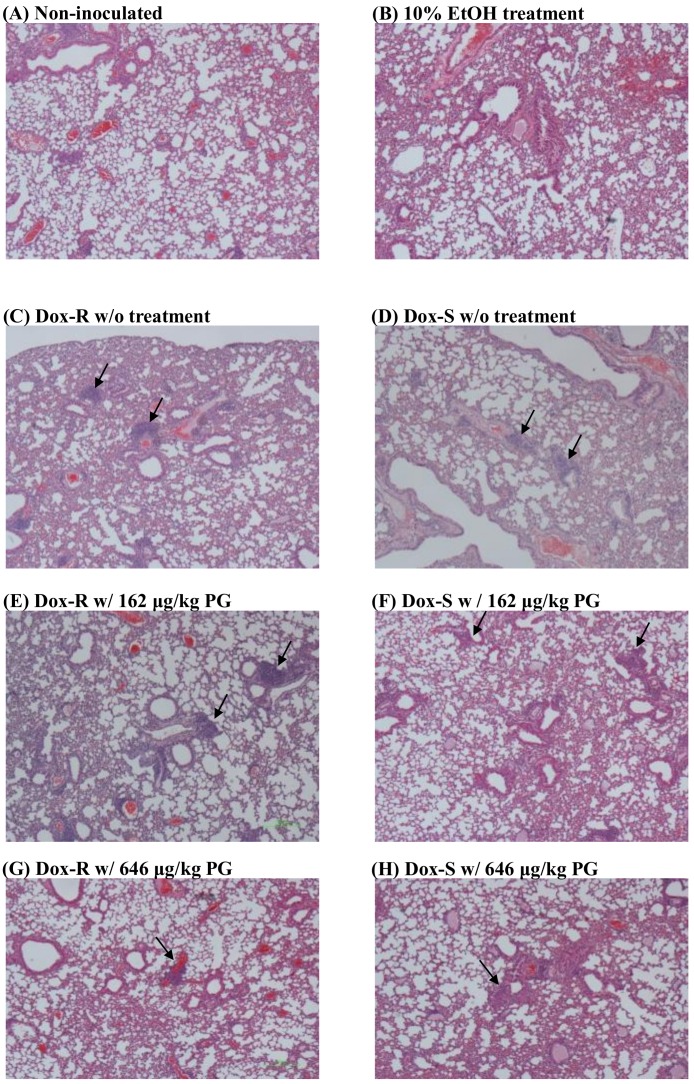
Figure 8.
In situ xenograft tumor after PG treatments. Lung tumor (A) without tumor injection, with tumor injection and treated with (B) 10% EtOH, (C,D) untreated, (E,F) 162 μg/kg PG, and (G,H) 646 μg/kg PG and stained with hematoxylin and eosin. All tissue snapshots were taken in 10 × 10. Black arrow points to the location of tumor cells.

