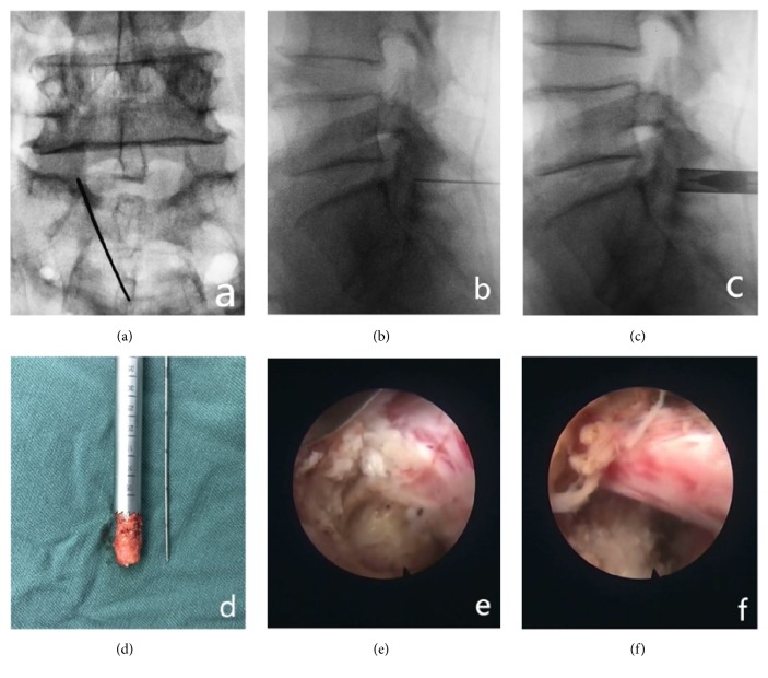
Figure 3.
(a), (b) Accurate positioning of lateral crypt stenosis with the following articular process in the center. (c) Part of the lower articular process is removed with a circular saw to establish a working channel. (d) Part of the lower articular process bone removed with a circular saw. (e) Herniated disc tissue is exposed outside the shoulder of the nerve root and removed. At the same time, the posterior edge of the vertebral body is removed, and the nerve root is decompressed. (f) The nerve root is dorsally, ventrally, and laterally decompressed thoroughly, allowing it to relax.

