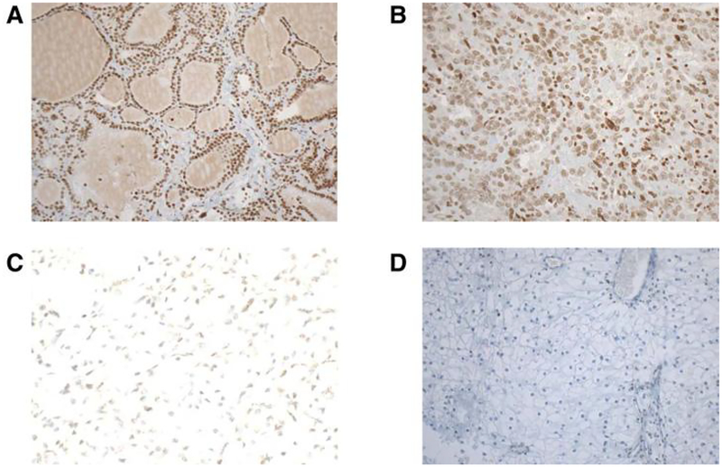
Figure 5.
Representative images of IHC staining for NSDΊ protein on whole slides of ccRCCs. A and B, Microphotographs are from a ccRCC sample positive for NSDΊ expression. A, Original magnification, ×ΊO. B, Original magnification, ×20.C,Weakstaining in one ccRCC case with NSDΊ promoter methylation. Original magnification, ×20. D, Negative staining in one ccRCC case with NSDΊ promoter methylation. Original magnification, ×ΊO.

