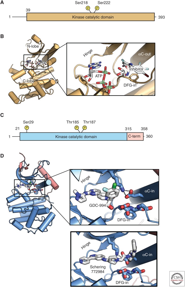
Figure 4.
Domain organization and structure of mitogen-activated protein kinase (MEK) and extracellular signal-related kinase (ERK). (A) Domain organization of MEK1. (B) Structure of the MEK kinase domain (PDB 1S9J). Inset shows features of interest, including the adenosine triphosphate (ATP) site, allosteric inhibitor site, and the αC-helix that adopts an “out” conformation, including disruption of the catalytic salt bridge. (C) Domain structure of ERK. (D) Structure of ERK kinase bound to GDC-0994 (blue with additions to the canonical kinase domain in red; PDB 5K4I). C-term, Carboxy terminal.

