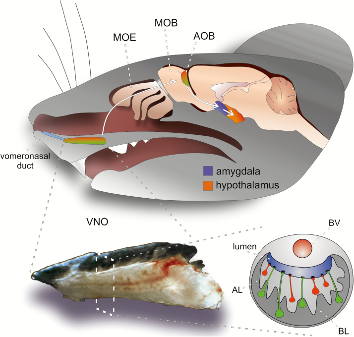
Figure 1.
Schematic overview of the mouse AOS. Shown is a sagittal view of a mouse head indicating the locations of the two major olfactory subsystems, including 1) main olfactory epithelium (MOE) and main olfactory bulb (MOB), as well as 2) the vomeronasal organ (VNO) and accessory olfactory bulb (AOB). Not shown are the septal organ and Grueneberg ganglion. The MOE lines the dorsolateral surface of the endoturbinates inside the nasal cavity. The VNO is built of two bilaterally symmetrical blind-ended tubes at the anterior base of the nasal septum, which are connected to the nasal cavity by the vomeronasal duct. Apical (red) and basal (green) VSNs project their axons to glomeruli located in the anterior (red) or posterior (green) aspect of the AOB, respectively. AOB output neurons (mitral cells) project to the vomeronasal amygdala (blue), from which connections exist to hypothalamic neuroendocrine centers (orange). The VNO resides inside a cartilaginous capsule that also encloses a large lateral blood vessel (BV), which acts as a pump to allow stimulus entry into the VNO lumen following vascular contractions (see main text). In the diagram of a coronal VNO section, the organizational dichotomy of the crescent-shaped sensory epithelium into an “apical” layer (AL) and a “basal” layer (BL) becomes apparent.

