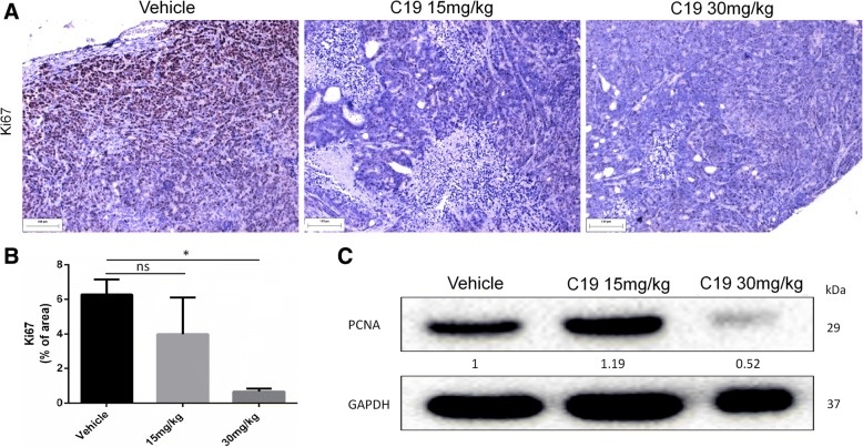
Fig. 6.
C19 treatment inhibits cell proliferation in vivo. a Tumor tissues from LoVo xenografts were evaluated by immunohistochemistry for Ki67 (a marker of proliferation). b The graph shows signal intensities of positive cells calculated using Image Pro Plus software. n = 3 *p < 0.05. c Tumor lysates were analyzed by western blot for expression levels of proliferating cell nuclear antigen (PCNA). Densitometric analysis was performed with Image Lab 4.0

