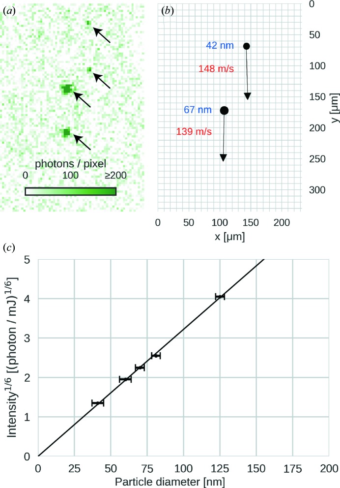
Figure 2.
Quantitative analysis of Rayleigh-scattering-microscopy data. (a) Double-exposure image of two polystyrene spheres of different diameters. The pulse delay was 50.8 µs and the pulse energy 56.1 mJ. (b) Extracted particle positions, velocities and diameters from the image shown in (a). (c) The sixth root of the mean integrated scattering intensity per particle (rescaled to 1 mJ laser-pulse energy) is plotted against the diameter of the respective polystyrene-sphere size standard. The values follow Rayleigh’s scattering law (solid line).

