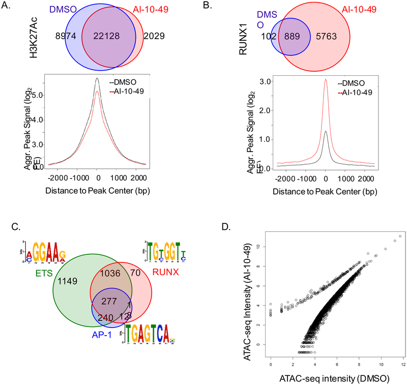
Figure 4. Global modification of RUNX1 binding to chromatin in inv(16) AML cells.
(A, B) Venn diagram of peak distribution (top) and aggregated peak signal from peak center (bottom) for H3K27ac (A) and RUNX1 (B) ChIP-seq peaks in ME-1 cells treated with DMSO (black) or AI-10–49 (red). (C) Motif analysis of RUNX1 associated peaks genome wide in AI-10–49 treated ME-1 cells. (D) Scattered plot representing open chromatin peaks by ATAC-seq analysis in DMSO and AI-10–49 treated cells. See also Figure S4.

