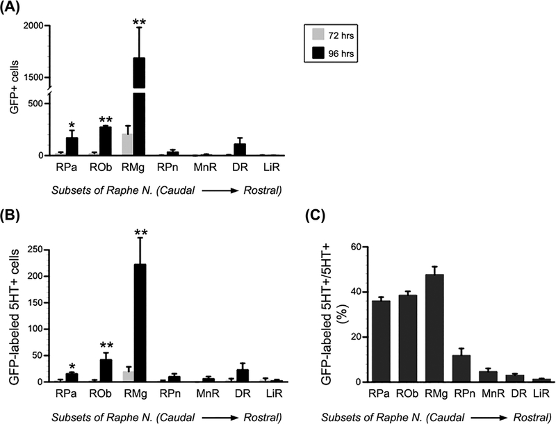
FIGURE 3.
GFP-labeled or GFP/serotonin (5-HT) double-labeled neurons are quantified in each subset of the raphe nuclei. A and B, Statistical analysis indicates significantly more (A) GFP-labeled or (B) GFP/5-HT double-labeled neurons in the caudal raphe nuclei at 96-hr post-infection than 72 h, including the RPa, ROb, and RMg. However, very few GFP-labeled or GFP/5-HT double-labeled neurons were detected in the rostral raphe nuclei, that is the RPn, MnR, DR, and LiR, at either 72 or 96 h post-infection. C, High percentage of GFP-labeled 5-HT+ to the total of 5-HT+ neurons is detected in the 3 caudal raphe nuclei (Unpaired Student’s t-test, *P < 0.05, **P < 0.01; RPa, raphe pallidus; ROb, raphe obscurus; RMg, raphe magnus; RPn, raphe pontis; MnR, median raphe, DR, dorsal raphe; LiR, linear raphe)

