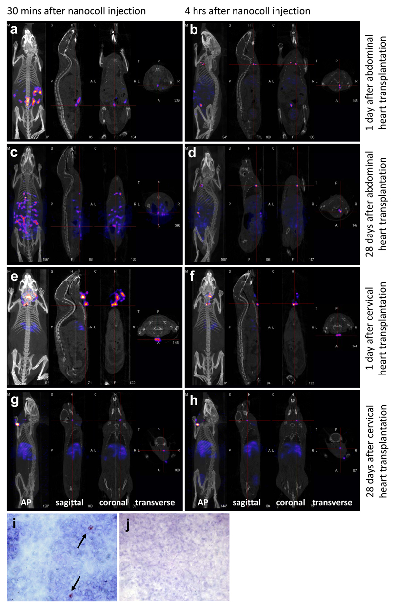
Figure 1.
a-h: SPECT/CT images taken after injection of nanocoll into the wall of the transplanted hearts. Representative images from one mouse are shown (n=3/4 per group). Each show images taken in 4 planes – (from left to right) the anterior posterior view, and images of the sagittal, coronal and transverse planes taken at the point of the red cross. Green arrows indicate the transplanted heart, and yellow arrows indicate the draining lymph nodes. a,b: one day after abdominal heart transplantation, the animal was scanned 30 minutes (a) and 4 hours (b) after nanocoll injection. c,d: 28 days after abdominal heart transplantation, the animal was scanned 30 minutes (c) and 4 hours (d) after nanocoll injection. e,f: one day after cervical heart transplantation, the animal was scanned 30 minutes (e) and 4 hours (f) after nanocoll injection. g,h: 28 days after cervical heart transplantation, the animal was scanned 30 minutes (g) and 4 hours (h) after nanocoll injection. All views shown are as labelled, except where oblique (b), and lateral (g, h) views are shown instead of AP to demonstrate the draining lymph nodes. Immunohistochemical staining for the donor MHC class II molecules I-Ad on the right anterior inferior mediastinal lymph node (i) and an axillary lymph node (j), harvested 8 days after abdominal transplantation of a DBA/2 (H-2d) heart into a C57BL/6 (H-2b) recipient. Representative examples of 4 independent experiments are shown. Black arrows indicate positive staining. Original magnifications x1000.

