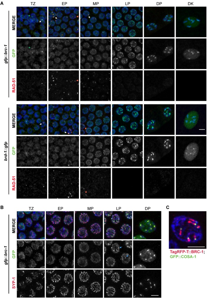
Fig 2. GFP::BRC-1 and BRD-1::GFP localize to the SC in meiotic prophase.
A) Nuclei from indicated meiotic stages stained with RAD-51 antibodies (red), DAPI (blue) and imaged for GFP fluorescence (green). White arrows demark foci positive for both GFP fluorescence and RAD-51 signal; green arrows demark foci containing GFP but not RAD-51; red arrows demark foci containing only RAD-51. Scale bar = 5 μm. B) Co-localization between GFP::BRC-1 (green) and SC central component SYP-1 (red) by antibody staining; germ lines at indicated stages were counterstained with DAPI. Blue arrows at late pachytene show chromosomal regions where GFP::BRC-1 concentrates before SYP-1. Scale bar = 2 μm. C) TagRFP-T::BRC-1 (red) and GFP::COSA-1 (green) at late pachytene showing TagRFP-T::BRC-1 on one side of the GFP::COSA-1 focus, which marks the persumptive crossover. Scale bar = 2 μm. Images are projections through half of the gonad. TZ = transition zone; EP = early pachytene; MP = mid pachytene; LP = late pachytene; DP = diplotene; DK = diakinesis.

