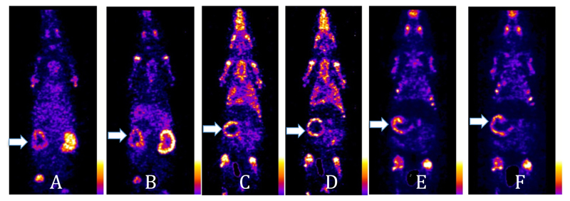
Figure 10.
A, B: Coronal sections of a rat scanned 30 min and 4 h, respectively after i.v. injection of [99mTc]-7. C, D: Coronal sections of a rat scanned at 30 min and 4 h, respectively, after i.v. injection of [99mTc]-4. E, F: Coronal sections of a rat scanned at 30 min and 4 h, respectively, after i.v. injection of 99mTc-1. Arrows show the uptake of the radiotracers in the calcified mesenteric arteries.

