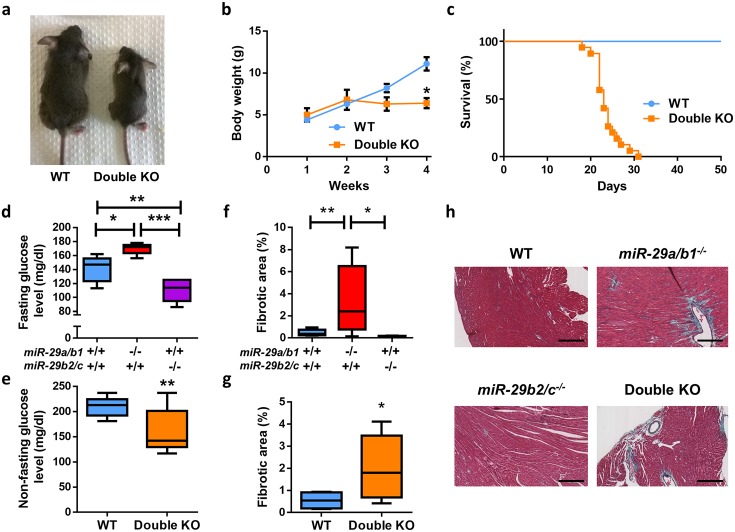
Fig 2. Cardiometabolic alterations of miR-29–deficient mice.
(A) Representative picture of 22-day-old wild-type and double KO mice. (B) Body weight curves of wild-type (n = 15) and double KO (n = 3) mice (p < 0.05 at 4 weeks, two-tailed multiple Student t test, Bonferroni-corrected). (C) Kaplan–Meier survival plot of wild-type (n = 11) and double KO (n = 19) mice (p < 0.0001; log-rank/Mantel-Cox test). (D) Fasting blood glucose level in wild-type (n = 10), miR-29a/b1−/− (n = 5), and miR-29b2/c−/− (n = 6) mice. (E) Nonfasting blood glucose levels in wild-type (n = 9) and double KO (n = 12) mice (two-tailed Student t test with Welch’s correction). (F) Percentage of collagen present in cardiac sections of wild-type (n = 12), miR-29a/b1−/− (n = 8), and miR-29b2/c−/− (n = 5) mice. (G) Percentage of collagen present in cardiac sections of wild-type (n = 4) and double KO (n = 10) mice (two-tailed Student t test with Welch’s correction). (H) Gomori’s trichrome stained heart sections of wild-type, miR-29a/b1−/−, miR-29b2/c−/−, and double KO mice (original magnification: ×10, scale bar: 200 μm). Original raw data can be found in S1 Data file. KO, knock-out; WT, wild-type.

