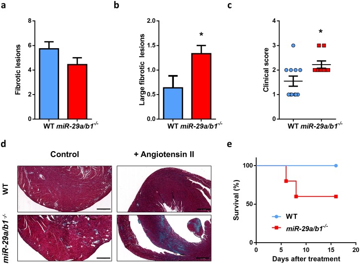
Fig 3. miR-29a/b1−/− mice display increased susceptibility to angiotensin II–induced cardiac fibrosis.
(A) Median of fibrotic lesions, (B) large fibrotic lesions, and (C) clinical score (grade 0: no fibrotic areas; grade 1: less than 25%; grade 2: from 26% to 50%; grade 3: from 51% to 75%; and grade 4: more than 76% of myocardium affected) in 3-month-old wild-type (n = 11) and miR-29a/b1−/− (n = 9) mice. (D) Representative micrographs of wild-type and miR-29a/b1−/− mice treated with angiotensin-II for 6 days (Gomori’s trichrome staining, original magnification: ×4, scale bar: 500 μm). (E) Kaplan–Meier survival plot of wild-type (n = 5) and miR-29a/b1−/− (n = 5) mice treated with angiotensin-II for 6 days. Original raw data can be found in S1 Data file. WT, wild-type.

