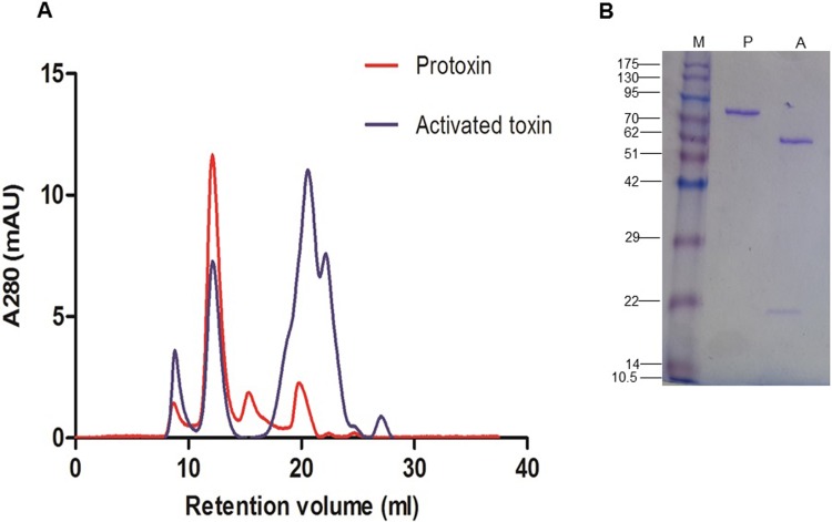
Fig 4.
Oligomeric structure of Vip3Aa65 protoxin and trypsin-activated toxin under native (A) and denaturing (B) conditions. A) Gel filtration chromatography in a Superdex 200 10/30 GL column. B) SDS-PAGE analysis of the 12 ml peak from the protoxin (P) and the activated toxin (A). M: Molecular weight markers in kDa.

