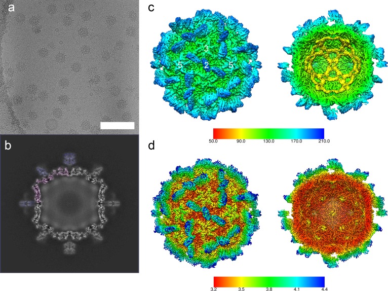
Fig 1. CryoEM and 3D reconstruction of MrNV VLPs.
CryoEM of MrNV VLPs revealed particles that were 40 nm in diameter with pronounced spikes on their outer surface (a, scale bar = 100 nm). A central section through the icosahedral reconstruction of the MrNV VLP reveals a sharply defined capsid shell (partially tinted pink) containing fuzzy density that we attribute to packaged nucleic acids. The P-domain spikes (partially tinted blue) were less well resolved in comparison to the shell, most likely a consequence of flexibility (b). An isosurface view of the reconstruction is shown coloured by radius and viewed along the icosahedral 2-fold symmetry axis (the symmetry axes are indicated by white numbers). A cutaway view reveals that the internal density forms a dodecahedral cage, consistent with that described for other nodaviruses (c). The sharpened map is also presented, coloured according to resolution, as both external and cutaway views (d). Sharpening of cryoEM maps down-weights lower-resolution information to reveal the fine structural details of the map, such as amino acid side chains. Poorly defined features such as the packaged nucleic acids are often lost upon sharpening because no high-resolution information is present. CryoEM, cryogenic electron microscopy; MrNV, M. rosenbergii nodavirus; P domain, protruding domain; 3D, three-dimensional; VLP, virus-like particle.

