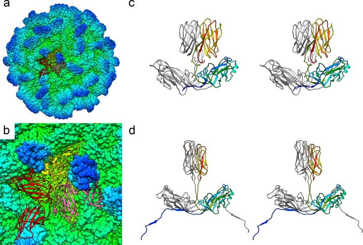
Fig 2. Atomic model of the MrNV capsid.
A solvent-excluded surface of the entire capsid is shown, coloured by radius (see Fig 1C for key). A single asymmetric unit comprising 3 copies of CP—CPA (red), CPB (yellow), and CPC (pink)—is shown as a ribbon diagram (a) and close-up view (b). Wall-eyed stereo-paired views of each dimer are shown (AB dimer [c], CC dimer [d]). In (c), CPB is presented in rainbow colour scheme, while CPA is coloured grey. In (d), one CPC chain is shown in rainbow colour scheme, while the other is grey. CP, capsid protein; MrNV, M. rosenbergii nodavirus.

