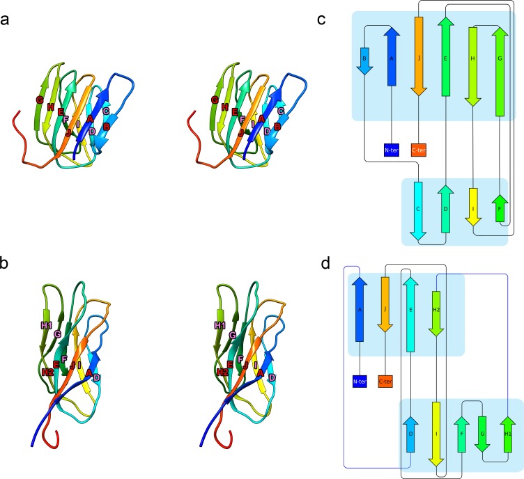
Fig 6. The MrNV P domain has a similar fold to that of the tombusvirus CNV.
A stereo-paired view of the CNV CPB P domain (PDB: 4LLF) is presented as a ribbon diagram with rainbow colouring (a). The diagram is annotated to indicate successive β-strands from the N- to C-termini that together make up a 10-stranded antiparallel β-barrel. A similar motif is seen in the MrNV P domain, which is likewise presented and annotated (b). Protein topology diagrams of CNV CPB P domain (c) and MrNV CPB P domain (d) present a simplified view to highlight the similarities of the P-domain folds in these 2 viruses. C-ter, C-terminus; CNV, cucumber necrosis virus; CP, capsid protein; MrNV, M. rosenbergii nodavirus; N-ter, N-terminus; P domain, protruding domain; PDB, Protein Data Bank.

