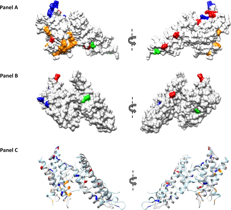
Fig 3. Mapping of SNPs on the structure of PvDBPII (Protein Data Bank code 4NUU) monomer and PvEBPII.
SNPs present specifically to Cambodian and Madagascar isolates are highlighted in red and blue respectively, whereas SNPs present in the isolates from both the countries are highlighted in green. Putative binding residues on PvDBPII are highlighted in orange. Molecular surface diagram of PvDBPII is shown. SNPs and putative binding residues of PvDBPII are highlighted (Panel A). Predicted structure of PvEBPII is shown as molecular surface diagram and SNPs are highlighted (Panel B). Helical ribbon representation of PvDBPII (in light blue) and PvEBPII (in light grey) are superimposed. SNPs and putative binding residues are highlighted (Panel C).

