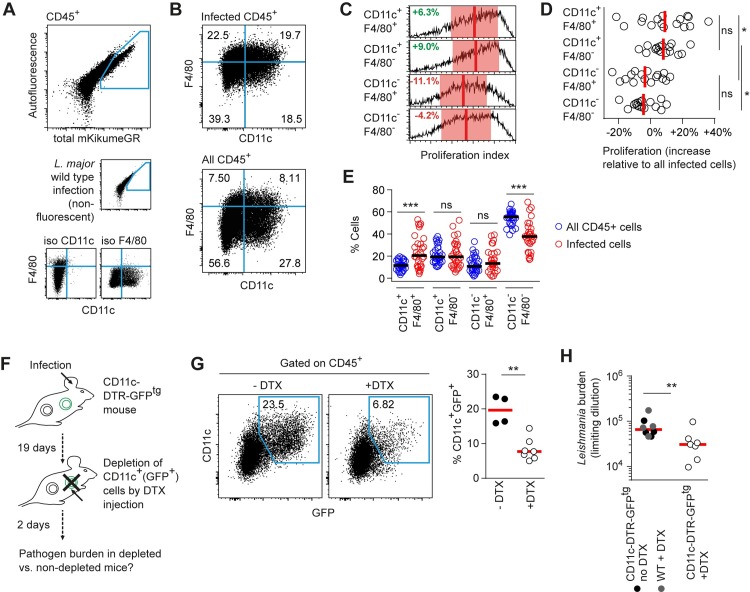
Fig 3. CD11c+ cells represent a niche for maintaining a high L. major burden.
(A-B) C57BL/6 mice ears were infected intradermally with LmSWITCH. 3 weeks post infection, parasites in the mouse ear were photoconverted and 48h later ears were harvested for flow cytometry analysis of intracellular L. major proliferation. (A) Strategy to identify L. major-infected cells (large plot) and CD11c+ and F4/80+ cell subsets (B) using controls infected with non-fluorescent L. major wildtype parasites (A, middle row, small plot) and isotype controls (A, lower row, small plots). (C) Comparison of proliferation rates of LmSWITCH located in the different cell compartments defined by F4/80 and CD11c. Vertical bars denote the mean, and shaded red boxes the standard deviation. (D) Quantitative analysis of the proliferation rates of LmSWITCH within the different cell populations shown in B-C. Each dot represents one mouse ear. Relative proliferation indices were obtained by normalization of the subpopulation’s proliferation index to the overall mean proliferation index within each sample. Vertical bars represent the mean. *p < 0.05; ns, not significant. Data pooled from three independent experiments. (E) Distribution of CD11c and F4/80 expression within infected (red symbols) and non-infected (blue symbols) CD45+ cells. Data shown are combined from the experiments shown in (D) and S2 Fig. Horizontal bars represent the mean. ***p < 0.001; ns, not significant. (F) Experimental approach for temporary Diphtheria toxin (DTX)-mediated CD11c+ cell depletion using the CD11c-DTR-GFPtg mouse model. (G) Quantification of the efficiency of depletion by measuring the percentages of GFP+CD11c+ cells in CD45+ cells isolated from infected mouse ears treated as shown in (F). (H) Limiting dilution analysis of L. major tissue burden in the ears of 3 week-infected CD11c-DTR-GFPtg mice, either PBS-injected (closed black symbols) or injected with DTX (open circles) 48h prior to analysis, or nontransgenic animals injected with DTX (closed grey symbols). **p<0.01. Each dot represents one individual mouse ear.

