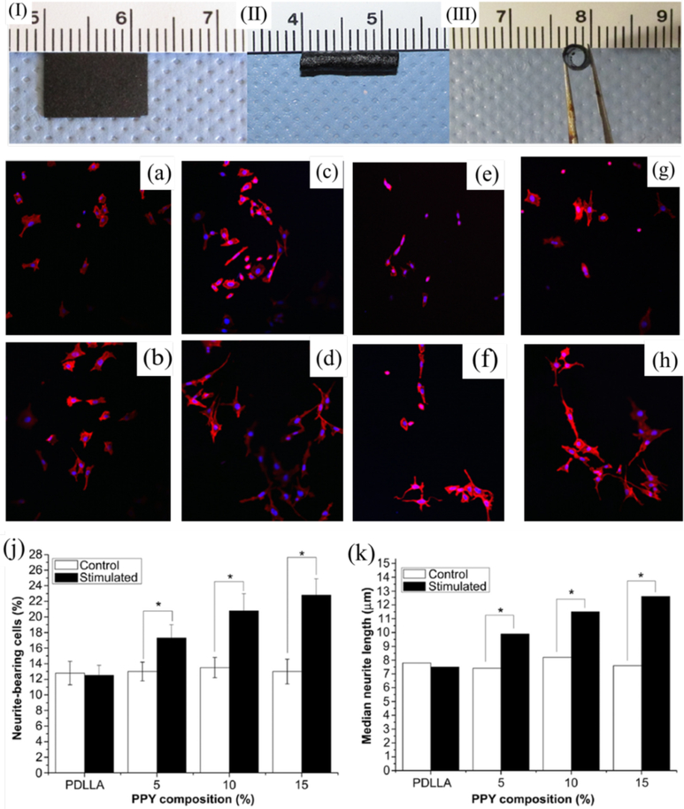
Figure 2.
Image of the PPY/PDLLA film (I) and the PPY/PDLLA nerve conduit (II, III). Fluorescent images of PC12 cells labeled for actin (red) and nuclei (blue). (a), (c), (e), and (g) are PDLLA, 5% PPY/PDLLA, 10% PPY/PDLLA, and 15% PPY/PDLLA without electrical stimulations. (b), (d), (f), and (h) are PDLLA, 5% PPY/PDLLA, 10% PPY/PDLLA, and 15% PPY/PDLLA with electrical stimulations of 100 mV for 2 h. Scale bar: 200 mm. (j) Percentage of neurite-bearing PC12 cells on conductive composite films with different PPY content (n= 4, *p < 0.05). (k) Median neurite length on PPY/PDLLA composite films with varying PPY composition. Reprinted from reference 53, Copyright (2013), with permission from Elsevier.

