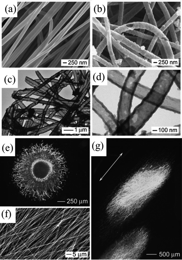
Figure 4.
SEM images of PCL nanofibers (a) and PPY nanotubes (b), respectively. TEM images of the PPY nanotubes (c, d). The nanotubes were prepared by soaking PCL/PPY core–sheath nanofibers in dichloromethane to selectively remove the PCL cores. Fluorescence micrograph showing the dorsal root ganglia (DRG) neurite field on random PCL/PPY core–sheath nanofibers (e), and aligned PCL/PPY core–sheath nanofibers (f). Fluorescence micrograph showing the DRG neurite field on aligned PCL/PPY core–sheath nanofibers (g). Reprinted with permission from reference 83. Copyright 2009 WILEY-VCH Verlag GmbH & Co. KGaA, Weinheim.

