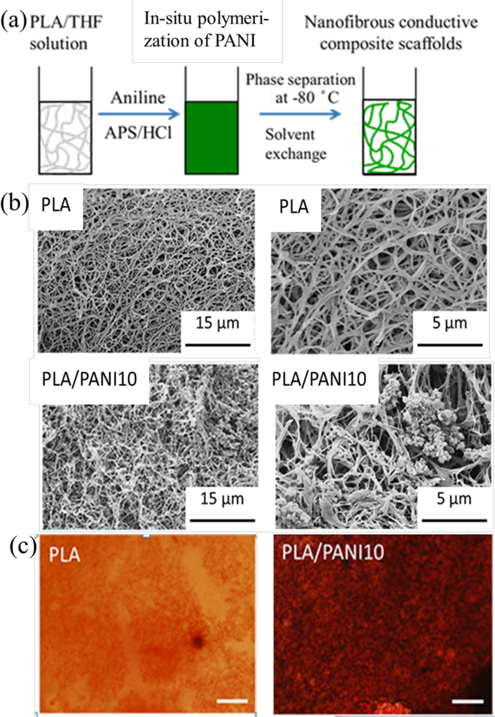
Figure 6.
(a) Schematic fabrication of conductive nanofibrous scaffolds. (b) SEM images of nanofibrous conductive scaffolds, PLA/PANI10: 10%wt PANI in the composite scaffolds, and the magnified images of scaffolds showing the nanofiber and PANI nanoparticles. (c) Alizarin red staining of BMSCs on different substrates for two weeks. Scale bar = 200 μm. Much larger area of positive alizarin red aggregates was present in the BMSCs of conductive composite scaffolds than that on PLA at day 14. Reprinted from reference 120, Copyright (2017), with permission from Elsevier.

