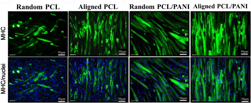
Figure 8.
Representative immunofluorescent images of myotubes differentiated for 5 days on random PCL fiber, aligned PCL fiber, random PCL/PANI3 fiber and aligned PCL/PANI3 fibers. MHC was immunostained green and nucleus blue. Reprinted from reference 86, Copyright (2012) Acta Materialia Inc., with permission from Elsevier.

