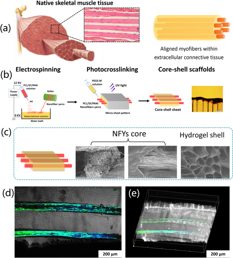
Figure 9.
(a) The composite core-shell scaffolds resemble the native structure of skeletal muscle tissue, consisting of aligned myofibers surrounded within extracellular connective tissue. (b) Preparation scheme of core-shell sheet scaffolds that mimic the native skeletal muscle tissue by the combination of aligned nanofiber yarns (NFYs) and hydrogel shell of poly(ethylene glycol)-co-poly(glycerol sebacate) (PEGS-M) solution. SEM images (c) of aligned NFYs core and hydrogel shell of the core-shell sheet scaffold after lyophilization. (d) The merged image of aligned NFYs cores exhibited overspreading C2C12 myotubes within the hydrogel sheet. (e) The 3D side view of the core-shell scaffolds containing highly organized myotubes. Reprinted with permission from reference 51. Copyright 2015 American Chemical Society.

