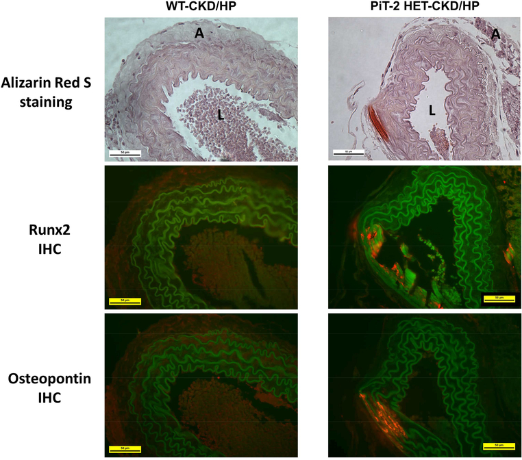
Figure 4 |. Immunohistochemistry of bone-related proteins in the abdominal aorta of CKD mice.
Sections of mouse abdominal aorta in the WT-CKD/HP and PiT-2 HET-CKD/HP groups were assessed by Alizarin Red S staining and immunohistochemistry for Runx2 and osteopontin. A, adventitia; CKD, chronic kidney disease; HP, high (1.5%) P diet; IHC, immunohistochemistry; L, lumen, P, phosphate; Runx2, runt-related transcription factor 2; WT, wild-type. Original magnification = 200x. Scale bar = 50 μm.

