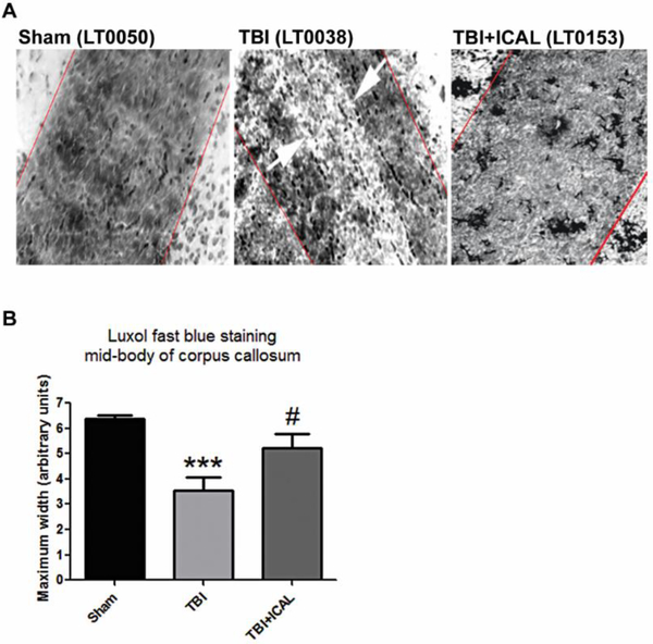
Figure 10. Pre-injury supplementation with Immunocal® improves axonal myelination of the corpus callosum in mice subjected to TBI.
A) Panels show Luxol fast blue-stained imaging of the mid-body of the corpus callosum at 20x magnification taken at 18 days post-TBI. Red lines indicate maximum width of mid-body and white arrows indicate area of demyelination observed in an untreated TBI mouse. B) Quantification of corpus callosum mid-body measurements. Untreated TBI mice displayed a statistically significant decrease in the maximum width of the corpus callosum mid-body when compared to Sham control mice (***p<0.001, n=5–7 mice per group; one-way ANOVA, p=0.001; effect size [95% confidence intervals] = −3.40 [−4.84 to −1.44]. Immunocal®-pretreated mice that were subjected to TBI showed a statistically significant increase in the maximum width of the corpus callosum midbody in comparison to untreated TBI mice (#p<0.05, n=5–6 mice per group; one-way ANOVA, p=0.001; effect size [95% confidence intervals] = 1.35 [−0.06 to 2.53]). Abbreviations used: ICAL, Immunocal®.

