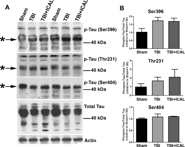
Figure 2. Western blotting for Tau phosphorylation and expression in brain tissue of mice subjected to TBI.
At 72h post-TBI, one half of the brain (excluding cerebellum) was dissected and homogenized in lysis buffer. Whole brain tissue lysates were resolved by SDS-PAGE and proteins transferred to PVDF membranes. A) Blots were sequentially stripped and re-probed with antibodies against Tau phosphorylated on Ser396, Thr231, and Ser404, total Tau, and actin (as a loading control). Asterisks indicate prominent phospho-Tau bands and MW markers are shown for estimation of size. The blots shown are from two independent sets of mice (Sham, TBI, TBI+ICAL) which displayed similar results. B) Densitometric analysis of each form of phospho-Tau was performed on three independent sets of mice. Phospho-Tau was normalized to total Tau and this value was set to 1.00 for each Sham mouse; the ratio of phospho-Tau to total Tau was then expressed relative to the Sham control for each set of mice. No statistically significant differences were observed; however, there was a trend towards increased Tau phosphorylation on Ser396 in untreated TBI mice compared to Sham and this trend persisted for TBI mice which had been pretreated with Immunocal® (one-way ANOVA, p=0.096). Abbreviations used: ICAL, Immunocal®; pTau, phospho-Tau.

