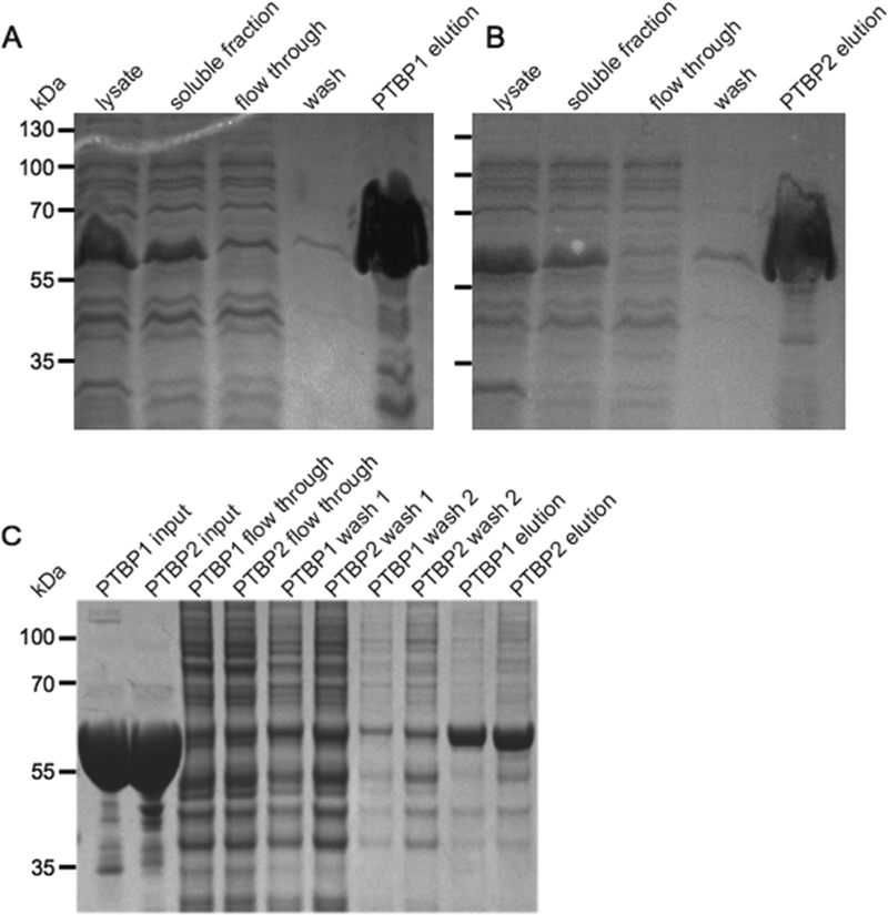
Figure 1.
(A and B) Purification of His6−tagged PTBP1 and PTBP2. Aliquots (10 μL) of the indicated fractions were analyzed by SDS− PAGE. The gel was stained with Coomassie blue dye. The positions and sizes (kilodaltons) of marker polypeptides are shown at the left. (C) Purification of His6−tagged PTBP from 50 μL splicing reaction mixtures containing HeLa nuclear extract, 2.2 mM MgCl2, 0.4 mM ATP, and 20 mM creatine phosphate. Aliquots (10 μL) of the indicated fractions were analyzed by SDS−PAGE. The gel was stained with Gel Code blue safe stain. The positions and sizes (kilodaltons) of marker polypeptides are shown at the left.

