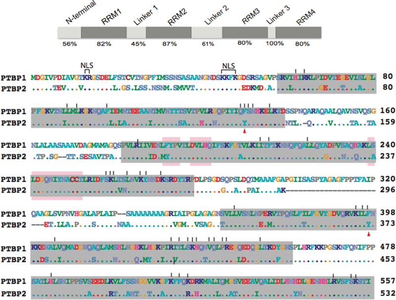
Figure 3.
Domain structure (top) of the PTB proteins indicating the segments defined in this study. The percent amino acid sequence identity between PTBP1 isoform 4 and PTBP2 is indicated below. Aligned amino acid sequences (bottom) of human PTBP1 isoform 4 and PTBP2. Gaps in the alignment are indicated as dashes. Residues identical to those of PTBP1 are shown as dots. RNA recognition motifs (RRMs) are highlighted in gray.38 Vertical lines above the sequence indicate PTBP1 residues that interact with RNA.38 Arrowheads below the sequence indicate RNA-interacting residues that are different in PTBP2. The bipartite nuclear localization sequence is indicated as NLS. PTBP1-Raver1-interacting motifs are marked with pink bars.

