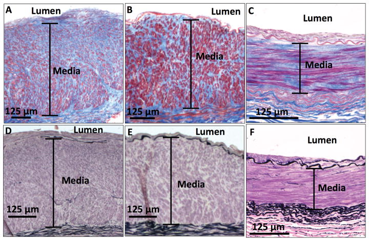
Figure 1. Medial fibrosis in the upper-extremity arteries.
Masson’s trichrome stain shows (A) an artery with severe medial fibrosis (86%) and (B) an artery with moderate medial fibrosis (65%) from patients with advanced CKD, as well as (C) an artery with representative medial fibrosis (50%) from a control subject. Elastic van Gieson stain of these samples shows that all arteries have very few elastic fibers in their respective medial layers (D, E, F). Collagen appears blue and smooth muscle cells appear red in Masson’s trichrome stain. Elastic fibers appear black in elastic van Gieson stain.

