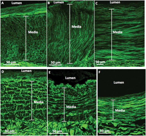
Figure 4. Representative second-harmonic-generation (SHG) images of the upper-extremity arteries and veins.
Upper panels: SHG images show (A) an artery whose medial collagen fibers had the honeycomb pattern and (B) an artery whose medial collagen fibers had the perpendicular to lumen pattern from patients with advanced CKD, as well as (C) an artery whose medial collagen fibers had the parallel to lumen pattern from a control subject. Lower panels: SHG images show (D) a vein whose medial collagen fibers had the railroad track pattern and (E) a vein whose medial collagen fibers had the parallel to lumen pattern from patients with advanced CKD, as well as (F) a vein whose medial collagen fibers had the parallel pattern from a control subject.

