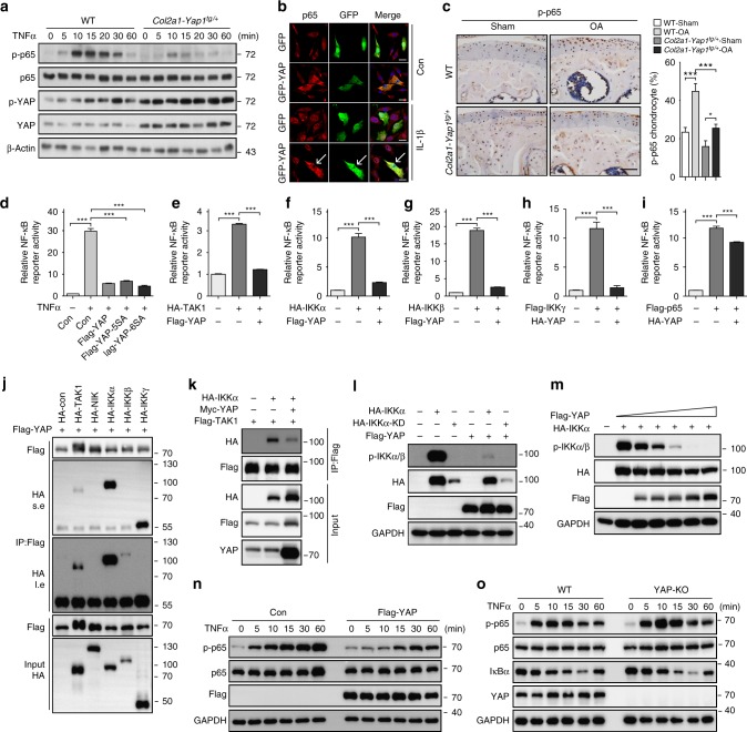
Fig. 7.
YAP attenuates NF-κB signaling by inhibiting IKKα/β activation. a Immunoblot analysis of primary chondrocytes with genotypes as shown after treatment with TNFα at 5 ng/ml. b Immunofluorescence analysis for p65 cellular localization in primary chondrocytes transfected with GFP-tagged YAP for 24 h and followed by treatment with IL-1β at 5 ng/ml for 1 h. Arrows indicate primary chondrocytes positively transfected with GFP-tagged YAP. c Immunohistochemistry of phospho-p65 expression in articular cartilage of mice 2 months after ACLT surgery with genotypes as shown. Scale bars, 50 μm. Statistical analysis of the percentage of p-p65 positive chondrocytes in articular cartilage of samples are shown in the right panel. d Luciferase assay of NF-κB luciferase reporter in HEK293A cells transfected with YAP, YAP-5SA, or YAP-6SA plasmid followed by treatment with TNFα for 6 h before collecting cell lysate. e–i Luciferase assay of NF-κB luciferase reporter 24 h after transfection of YAP with respective NF-κB component plasmids as indicated in HEK293T cells. j Immunoprecipitation assay to detect the association of YAP with indicated kinases related to NF-κB signaling in HEK293T cells. s.e. short time exposure of film. l.e. long time exposure of film. k Immunoprecipitation assay of IKKα and TAK1 with or without overexpression YAP in HEK293T cells. l Immunoblot analysis of the phosphorylation of IKKα with overexpression of YAP in HEK293T cells. m Immunoblot analysis of the phosphorylation of IKKα with expression of different dose of YAP in HEK293T cells. n Western blot analysis of lysate of HEK293A cells transfected with Flag-tagged YAP after treatment with TNFα as indicated time. o Immunoblot analysis of the phosphorylation of endogenous p65 after treatment with TNFα as indicated time in WT or YAP-KO HEK293T cells. All data are presented as mean ± SD. *p < 0.05, **p < 0.01, ***p < 0.001. One-way ANOVA followed by Tukey’s test was performed. All experiments were repeated three times independently

