
Figure 5.
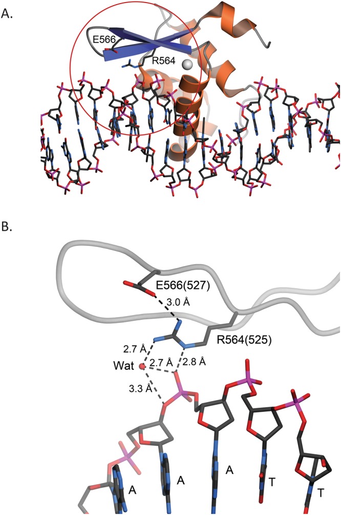
Residue R525 of human FOXP1 is predicted to interact with both DNA and nearby residues. (A) Chain K of the forkhead domain of human FOXP2 (PDB 2A07) bound to DNA43. DNA is depicted in ball-and-stick and FoxP2 is shown as a ribbon diagram with α-helices colored orange and β-strands colored blue. The forkhead domain of FoxP2 shares 87% sequence identity with human FoxP1. Residue R564 in FOXP2 (corresponding to R525 in FOXP1) is located at the protein-DNA interface. The red circle shows the area that is depicted in panel B. (B) A closer view of the environment of R564 (R525 in human FOXP1). Residue numbers in PDB 2A07 are indicated; their corresponding human FOXP1 numbering shown in parentheses. Potential hydrogen bonding interactions are shown in dotted lines with distances indicated in Ångstroms. “Wat” indicates an ordered water molecule mediating H-bonds between protein and DNA. R525 in human FOXP1 is likely to make similar contacts, which the R525Q mutation likely disrupts. Figure made with POVScript+47.