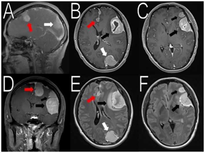
Figure 1.
Initial imaging showing multiple menigniomas. Preoperative contrasted T1 and T2 MRI scans reveal multiple meningiomas. The left calcified parafalcine lobulated mass (2 × 2.2 × 3.3 cm, AP, TV, CC) was associated with vasogenic edema (red arrows in A,B, D–E). In the left frontal temporal convexity, there was a 4.5 × 2.9 × 4.1 cm (AP, TV, CC) mass with another 2.3 × 2.0 × 1.9 cm (AP, TV, CC) mass located superior to it (black arrows in B–F). In the occipital lobe, the mass was measured to be 2.6 × 2.9 × 3.9 cm (AP, TV, CC) (white arrows in A,B,E). There was an 8 mm rightward midline shift. The images seemed to suggest neurofibromatosis type 2.

