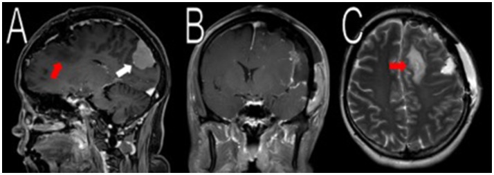
Figure 2.
Resection of the frontal/parietal/temporal mass. Post-operative MRI contrasted T1 and T2 scans showed resection of the meningiomas in the left frontal/parietal/temporal convexity with expected post-operative changes (red arrows in A,C, not shown in B). The occipital lobe mass was visible from the sagittal view (white arrow in A).

