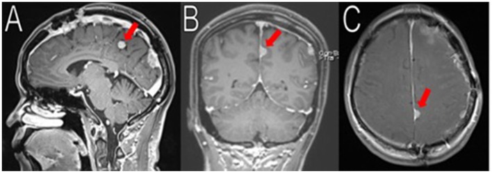
Figure 5.
Latest imaging. Her most recent MRIs (16 months after her last surgery) show multiple enhancing extra-axial masses, stable compared to her immediate post-operative MRIs. Here is a stable 1.8 cm (superior-inferior) meningioma arising from the left posterior falx, adjacent to the prior resection cavity (red arrows in A–C). No recurrence observed.

