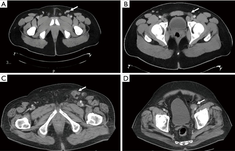
Figure 1.
CT scans demonstrating a false-positive and true-positive when staging ILN. A 32-year-old man with HgT2 penile cancer invading into the glans with imaging demonstrating a 2.6 cm left inguinal node (arrow) (A) and 1.9 cm left external iliac node (arrow) (B) all of which were benign after robotic-assisted node dissection. An 82-year-old man with HgT2 penile cancer with 2.2 cm left inguinal node (arrow) (C) with irregular borders and 1.8 cm left external iliac node (arrow) (D). Inguinal lymph node dissection was performed with pathology demonstrating metastatic disease with tumor necrosis and extranodal extension.

